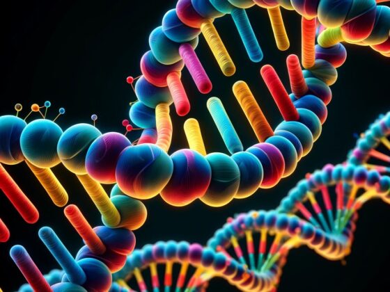
The Fluorescence In Situ Hybridization (FISH) test is a lab technique widely used in cytology to detect and locate specific DNA sequences on chromosomes. It is particularly useful in interpreting and making diagnosis of cancer and genetic disorders. Fluorescent deoxyribonucleic acid (DNA) probes, which are attached to the high degree of complementary parts of the chromosome, emit the colored signals. These signals could be grasped and visualized using fluorescent DNA probes.
The FISH Test Process:
Sample Preparation: Cells from a patient, such as a blood sample or tissue biopsy, are fixed onto a glass slide.
DNA Denaturation: The DNA within the cells on the slide is made single-stranded (usually by heating), which exposes the DNA sequence of interest making it accessible for binding.
Probe Design: A probe, which is a piece of DNA with a fluorescent label attached to it, is created to match the specific DNA sequence being investigated.
Hybridization: The fluorescent probe is applied to the slide with the sample. The probe searches for and binds (hybridizes) to its complementary DNA sequence on the chromosomes in the cells.
Washing: The slide is washed to remove any probes that haven’t bound to their target sequence, reducing background fluorescence.
Probe Detection: The slide is examined under a fluorescence microscope. The probe will emit light at a certain wavelength (color) when exposed to a laser, showing exactly where the target DNA sequence is on the chromosome.
Analysis: A specialist, often a cytogeneticist, analyzes the fluorescent signals to determine the presence, absence, or rearrangement of specific DNA sequences.